
|
|
|
| Home | Topics Research | Publications | Collaborations | Contact Us | People | Links |
|
|
Micro
CT


 CT analysis of Roman Tooth With collaboration of "Pigorini" Museum, Rome, Italy 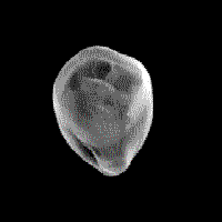 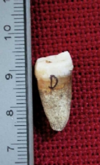
Human tooth with carie (found in the Isola Sacra necropolis) The specimen features: Dim: 2.0 x 1.0 x 0.7 cm3 V / I : 70 kVp / 1000 A ANGULAR STEPS: 600 VOXEL: 30 m DETECTOR: XCCD |
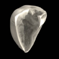
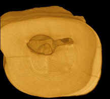
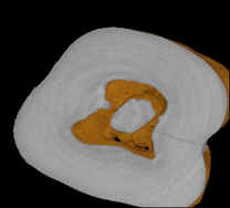
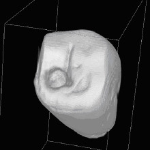 The micro-CT system shows its utility in the analysis of paleobiological samples. In collaboration with the Anthropology Section of Museo Nazionale L.Pigorini of Rome, a detailed program of micro-CT analysis has been set up for the investigation of paleoprimates. Figures show the picture of a human molar from the necropolis of Isola Sacra, near Rome. Tomography allows a 3D reconstruction of the tooth with a resolution of 30 μm (voxel side). Successively, through virtual cuts, parts of the volume can be removed to provide an inside view of the sample, enabling tooth lesions to be easily located. A specific diagnosis of caries can be made with certainty based on the results of this analysis. Micro-CT represents, today, the only method of analysis for this kind of sample since it offers an opportunity to obtain complex morphological information with high spatial resolution.In
Figures left: 3D reconstruction of the tooth. Virtual cut into the volume serving to locate the lesion.
The micro-CT system shows its utility in the analysis of paleobiological samples. In collaboration with the Anthropology Section of Museo Nazionale L.Pigorini of Rome, a detailed program of micro-CT analysis has been set up for the investigation of paleoprimates. Figures show the picture of a human molar from the necropolis of Isola Sacra, near Rome. Tomography allows a 3D reconstruction of the tooth with a resolution of 30 μm (voxel side). Successively, through virtual cuts, parts of the volume can be removed to provide an inside view of the sample, enabling tooth lesions to be easily located. A specific diagnosis of caries can be made with certainty based on the results of this analysis. Micro-CT represents, today, the only method of analysis for this kind of sample since it offers an opportunity to obtain complex morphological information with high spatial resolution.In
Figures left: 3D reconstruction of the tooth. Virtual cut into the volume serving to locate the lesion.
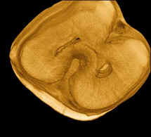
CT analysis of Pakistan Prehistoric Tooth (7,500-9,000 years ago)
With collaboration of "Pigorini" Museum, Rome, Italy 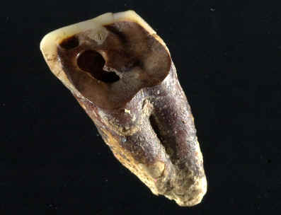
From Nature Paper: "Microtomography revealed cavity shapes that were conical, cylindrical or trapezoidal. They also showed concentric ridges preserved on some walls that had been left by the drilling tool. The teeth of at least one individual reveal that the procedure involved not just removal of the tooth structure by the drill, but also subsequent micro-tool carving of the cavity wall by either the operator or the patient. In all cases, marginal smoothing confirms that drilling was performed on a living person who continued to chew on the tooth surfaces after they had been drilled."
3D CT carried out at Synchrotron ELETTRA, Trieste, Italy:
Webmaster: Rosa
Brancaccio