
|
|
|
| Home | Topics Research | Publications | Collaborations | Contact Us | People | Links |
| IORT (Intra
Operative Radiation Therapy) Project |
AN ELECTRON BEAM IMAGING SYSTEM FOR QUALITY ASSURANCE IN I.O.R.T.
 |
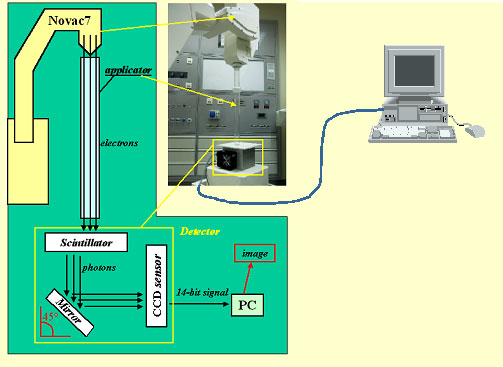
Intra Operative Radiation Therapy (I.O.R.T.) is a special radiotherapy technique based on delivery of a single high dose of ionising radiation to the cancerous tissue during oncological surgery.
Novac7, a new kind of linear accelerator (manufactured by Hitesys), has been expressly conceived for direct use in the surgical room. Novac7 can deliver electron beams of different energies (3, 5, 7 and 9 MeV), with a high dose rate (up to 20 Gy/minute).
These peculiar characteristics give rise to some complications with classical dosimetric techniques. |
|
THE ACQUISITION LINE |
 |
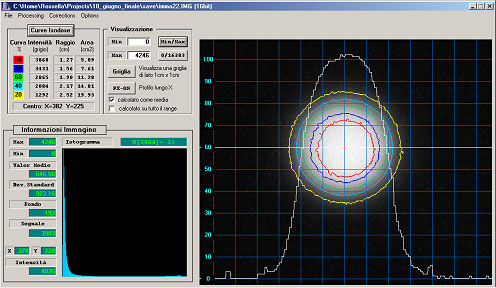 |
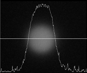
THE SOFTWARE The dedicated software is able to pilot the CCD camera and, within few seconds, to visualise a 14-bit digital image on the monitor. Moreover, it provides some useful information, such as: - electron beam position and size, - beam profiles for uniformity and symmetry evaluation - and isodose curves. |
|
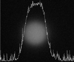
SPIKES CORRECTION |
  |
|
SYSTEM ANALYSIS |
 |
|
SYSTEM ANALYSIS |
 |
CONCLUSIONS
The system is able to obtain 2D measurements of dose distributions in real time.
The special frame grabber software gives several information on the radiation field properties: beam position and size, uniformity and symmetry, isodose curves. Our device can therefore be used for fast controls on the electron beam prior to treatment and for quality assurance on a regular basis.
Webmaster: Rosa
Brancaccio