
|
|
 |
| Home | Topics Research | Publications | Collaborations | Contact Us | People | Links |
Autoradiography
|
Autoradiography is a methodology
used for different applications, especially in the biological ones. Its aim is
to detect spatial distribution of a radioisotope and calculate its concentration
in a particular region of the sample. Since the beginning of the last century
autoradiography is known as a research method. After a long period characterized
by conventional techniques based on photographic principles and screen films, in
recent years it evolved towards digital acquisition, together with the
development of new materials as scintillators and semiconductors.
Today, conventional autoradiography is still the most used technique, because of
its practicality, lower costs and best resolution.The disadvantage related with
this traditional technique is the very long time requested for an acceptable
autoradiographic representation (up to several hours). The main task of the new
research in digital autoradiography is the analysis in presence of low
quantities of radioactivity and faster image acquisition time.
In particular this feature is very important in dynamic studies with
radiotracers in biological tissues. The opportunity to reach this goal is now
possible with the use of a new kind of image intensifier – the electron
bombarded CCD.
The aim of our work is to develop a system for digital autoradiography based on
this new device. Preliminary tests with our equipment have shown that real time
imaging with commonly used radiotracers is feasible.
|
|
 |
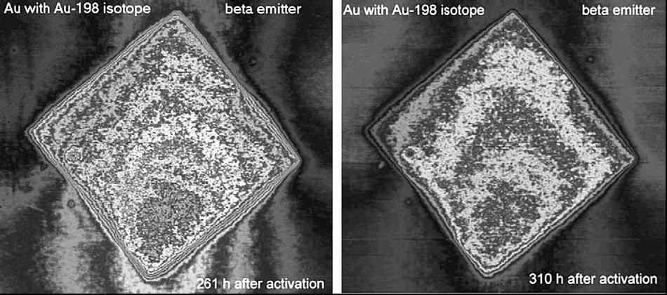
Images of the neutron activated sheet of gold taken in different dates.
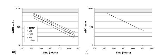
Decay curve measured at different positions on the sheet of gold.
Real time autoradiography
|
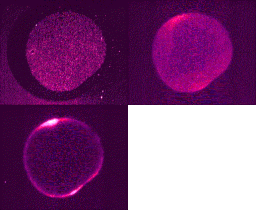
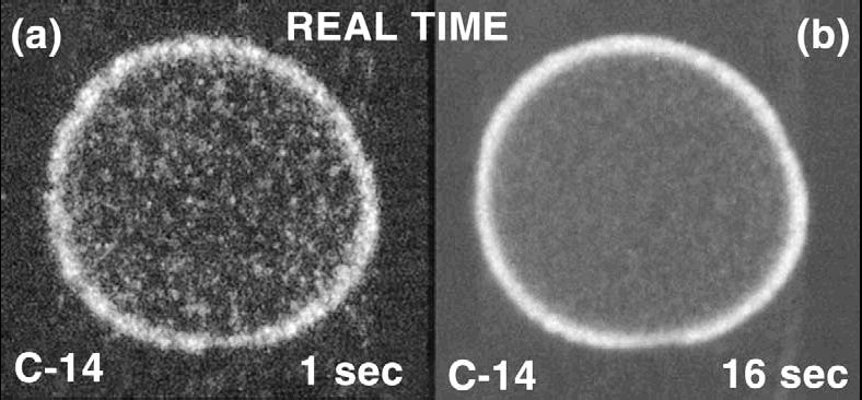
A very interesting result has been achieved with our system: real time autoradiography of carbon-14 labeled radiotracers has been made. This is mainly due to the system capability to detect single beta particles. In fact the detector has a very low noise and the weak signal of carbon-14 beta can be detected. It is possible to reveal the shape of a radiotracer drop put on a millipore filter in only about 1 s, including readout time (Fig. 5(a)). |
 |
 |
 |
| Sequence |
Animated sequence |
Webmaster: Rosa
Brancaccio