X-Ray Digital Radiography and Computed Tomography applied to Cultural Heritage, Medicine and Industrial applications
 
Home Topics Research Publications Collaborations Contact Us People Links
 

Linear Scanner for NDT
Digital Radiography
Computed Tomography
   Intensified Cone Beam CT
   Micro CT
   Multislice Tomography
   Works carried out at the Getty
       Conservation Institute, Los Angeles
   High Energy Tomography
  
Large Objects
      Ancient Globe
Neutron Tomography
Iort Project
Autoradiography

Large Objects Tomographyc System


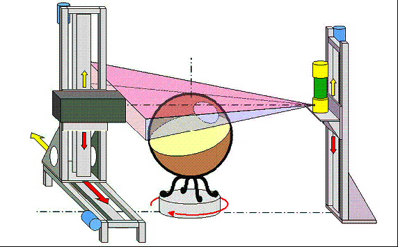
THE EXPERIMENTAL SETUP 
The CT system used consists of an X-ray tube, mounted on a vertical moving axis, a planar detector made of a GOS scintillating screen and an Electron Bombarded CCD (EBCCD) camera, which can translate both horizontally and vertically, and a rotating platform, on which the globe was placed. 

A motor moved the X-ray tube along an aluminum column. Two more motors moved the detector along the x and y-axis. The main problem of getting a 
complete CT is related to the large size of this masterpiece (220 cm in diameter) and the need of achieving an in situ analysis in a museum surrounded by visitors. The dimension of the planar detector used was 30 × 40 cm2, thus requiring the acquisition of about 32,000 images, for covering the entire Field Of View (FOV).


Webmaster: Rosa Brancaccio