|
|
 |
| Home | Topics Research | Publications | Collaborations | Contact Us | People | Links |
|
|
Multislice Tomography 


An innovative CCD-based high resolution CT system for analysis of trabecular bone tissue |
Synchrotron based digital radiography and microtomography devices are powerful, non-destructive, high-resolution research tools.
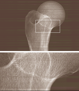
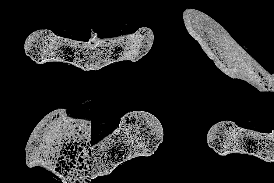
A high resolution computed tomographic system with 22.5 micrometers pixel size and FOV of 13 cm × 18 cm was set up for a synchrotron source. The possibility of analyzing a sample of large size permits the reconstruction of CT slices of big parts of bones or even complete bones (i.e., proximal femur, wrist), leaving them intact and without special sample preparation (i.e. specimen cutting, extraction). We demonstrated the feasibility of using such images to extract information about the trabecular structure by scanning the proximal part of a human dried femur, containing greater trochanter, femoral neck and femoral head.
 |
 |
|
3D Reconstruction |
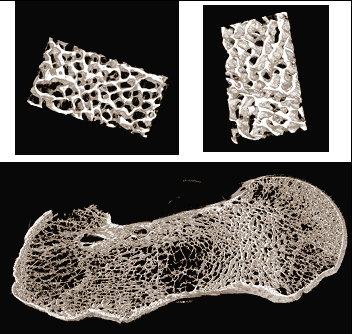
Three-dimensional images obtained from a stack of 50 microCT slices of the examined human femur. Top row: two particulars of an ROI. Bottom row: reconstruction of the entire cross sectional area. |
 This system is an improvement with respect to standard microCT systems, since it gives the possibility to have data on a complete bone and not just in a limited ROI. The spatial limits (specimen size) were overcome in the present system because of the use of a special fiber optic adapter. The linear pixel size used (22.5 micrometers) is optimal for the imaging of trabecular bone structure.
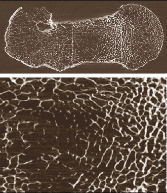
The quality of the reconstructed cross section images confirms that this investigation technique permits high-resolution three-dimensional imaging of large volumes (up to 18 cm × 13 cm × 13 cm) at a pixel size not achievable with conventional microCT scanners.
In the near future, we are planning to test the system with regular X-ray microfocus tubes, in order to assess the practicability of performing in-vivo imaging of human patients.
We would thank the staff of the SYRMEP beamline at ELETTRA Synchrotron Light Laboratory, in particular Diego Dreossi and Silvia Pani. Authors are also grateful to the ‘NECTAR CT system’ and CEFLA group (Eros Nanni, Andrea Albertini and Susanna Del Nero).
Figures (left): Reconstructed tomographic slice of the human femur: the trabecular structure is clearly visible over the entire FOV. Top: entire FOV. Bottom: enlargement of the box shown above.
Webmaster: Rosa
Brancaccio