
|
|
|
| Home | Topics Research | Publications | Collaborations | Contact Us | People | Links |
|
|
Multislice Tomography


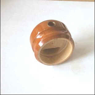  CT analysis of an Ancient Flute In collaboration with "Accademia Filarmonica di Bologna" |
The tomographic analysis of two samples, an ancient flute and a fossilized Oreopithecus jaw, was carried out in order to evaluate the performance of the detector for CT. Both radiographic data-sets consist of 720 radiographic projections over 360° with an angular sampling of 0.5°. The X-ray tube was set with a tungsten anode at 100 kV with an aluminium filter thickness up to 3 mm in order to reduce beam-hardening artifacts. At each angular position, the linear array simultaneously acquires the transmission values for 35 slices. The CT slices are reconstructed using the Feldkamp algorithm.
The voxel size depends on the magnification factor. These tests show the efficiency of this system on either small or large objects. It is possible, using the same camera, without any significant changes, to take digital radiographies of features of various dimensions. CT reconstructions of small objects (i.e. Oreopithecus dimension: 3 x 5 x 2 cm3), obtained from the EBCCD system, were compared with reconstructions obtained using a micro-CT system.
Results show no particular differences between the two reconstructed slices. We therefore tested CT reconstructions of larger objects with similar nominal spatial resolution. A multi-slice of an ancient flute (diameter = 6.7 cm) was reconstructed with a voxel size of 13.9 microns. The described detector has the following advantages over other linear multi-slice detectors:
1) the samples treated can reach dimensions in the order of 100 mm compared to the dimensions of a few mm for other systems with the same spatial resolution;
2) by changing the radiation-light converter, it is possible to change the energy of the X-rays or to perform high resolution neutron radiography;
3) if all the electronics is outside the beam no damage is caused by radiation, a positive factor for high energy X-rays or neutron beams;
4) as the light is detected by an EBCCD, a low dose is required for taking images, which means low currents for the X-ray tube and, consequently, a small focal spot. With regards to the systems drawbacks, we would like to point out the imperfect contact between two adjacent bundles, (gap in the order of 0.1 mm), which requires correction by software during the image reconstruction phase.
Webmaster: Rosa
Brancaccio