|
|
|
| Home | Topics Research | Publications | Collaborations | Contact Us | People | Links |
|
|
Intensified Cone Beam CT |
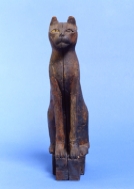
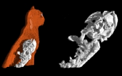
This detection system has been used to investigate small Egyptian mummies preserved in the archaeological Museum of Bologna. A careful analysis was carried out on a cat-shapedcoffin (upper picture). The size of the sarcophagus is 37.7×10×19.5 cm3 and it has a structure composed of different materials. DR and CT can help to determine the position and characteristics of the mummy inside the coffin. CT data allow very fine distinctions to be made among materials with different densities, thus providing a large amount of information. Figures (down) show a 3D reconstruction of the sarcophagus and how it is possible to extract the cats skeleton for a detailed analysis of the mummy.
Webmaster: Rosa
Brancaccio

 Small Egyptian cat-shaped coffin