|
|
|
| Home | Topics Research | Publications | Collaborations | Contact Us | People | Links |
|
|
Intensified
Cone Beam CT |
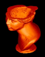
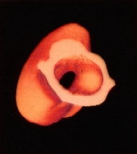
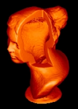
An experimental 3D X-ray CT system for industrial Non Destructive Evaluation (NDE) applied to small specimens (< 20 cm) has been set up at the Physics Department of University of Bologna (Italy). Cultural heritages present a wide set of objects that can be classified by material composition, size and archaeological interest. As regards of tomographic applications, sculptures represent the most important samples to be inspected because they are generally inaccessible inside and information about their integrity can be retrieved only for the outer parts. Moreover, materials used for sculptures can be classified into three classes only: metals, stones (limestone and argil composites - marbles) and woods. The inspection of metals requires high energy CT system, whereas sculptures of wood and stone of limited size can be investigated by conventional radiographic tubes used in medical systems. Cone beam CT analysis was carried out on a small argil bust from Pompei, a hand-made reproduction of a larger statue representing an ancient divinity. The data-set consists of 120 projections at 3° intervals over 360°, as required by Feldkamps algorithm; the total data acquisition time was of the order of 1 ½ hours. The object was irradiated only on the upper part, limiting the radiographic image to an area of 9x7 cm2. Magnification factor is about 1. Fully 3D visualization of a sample provides the restorer with a easier straightforward way of visualizing internal structures.
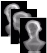
Radiographyc Sequence
Virtual cuts:
Webmaster: Rosa
Brancaccio
 Pompeian Head